Medical laboratory workshop Episode 10: Diagnosis and identification of Plasmodium falciparum using stained blood film
Welcome once again to another edition of our workshop series where we expose pathogens behind various disease of human importance. Today's discussion is about what is endemic in the tropics. As usual, we give a brief overview about this insects before going deep into discussing their clinical significance.
Introduction
Mosquitoes remain a threat to human existence and till date, efforts are being made everyday to curtail their effects. Those of Medical importance belong to the sub family Culicinae which is further divided into three tribes - Anophelini, Culicini (Aedes, Culex and Mansonia) and Megarhini (the giant mosquito).
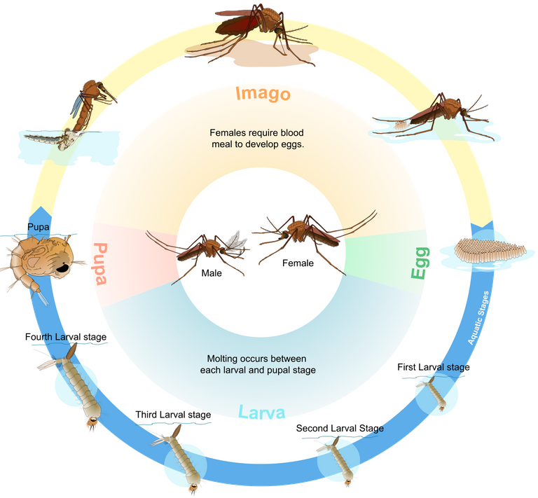
Mosquito life cycle
They are small slender bodily flies with proboscis (a forward projecting structure for obtaining blood meal). They have different sexes, the males suck nectars of flowers and do not pose any threat to humans, they hardly live for more than a week and their main role is to fertilize the females.
These males die after fertilizing the female mosquitoes while the female ones especially the female anopheles sucks blood from humans because they need this blood for the maturation of their fertilized egg. After the females lay their eggs, they also die off. The females remain quiescent for about 2-3 days when they are fully fed, for the maturation of their ovary, they remain indoors where they get their blood meal from humans, while when outdoor they rest in sheltered places like under surface of leaf, caves, or cans patiently waiting for preys.
After the eggs are laid (usually in hundreds) they may hatch within 24hours depending on the availability of water. That is to say that, the eggs need water to hatch. This explains why during rainy season, you tend to have numerous mosquitoes flying around. If water is not available, these eggs can remain unhatched for weeks or months.
Once the eggs are hatched, the larva are released, these larvae have a stiff hairs and breathing trumpet-like siphon which they use to pierce the surface film of water for gaseous exchange, and being aquatic wrigglers (they are mostly found in water where they breath and feed on microscopic organism) they undergo further development into the pupa stage which resembles a lobster and finally transforms into the adults.
On a side note, simple way to eliminate them at this stage is to either introduce Gambusia affinis - the mosquito eating fish into the pond or any stagnant water, so that they can feed on the mosquito larvae or you pour oil on the surface of the water. This will suffocate and prevent them from breathing and ultimately leading to death.
These final transformation can take 2days to a week depending on the temperature and they can suck blood within twenty four hours after maturation into adult.
Mosquitoes mostly bite during the night and are usually active between the late hours of the day from 6:30pm to early morning, 7am and are able to detect and suck blood from humans basically because of four main reasons - our smell, the carbon dioxide we release, the heat we generate, and the lactic acid produced on our skin.
Once they locate the perfect spot ok their host, they use their proboscis to secrete saliva on the skin, this saliva contains an anticoagulant (prevents the blood from coagulating or clotting), some vasodilators that keeps the vein dilated so that blood keeps flowing to the region or spot where they are actively obtaining blood. The saliva also contains a chemical that acts like an anaesthetic to make you unaware of its bite most times.
The female anopheles mosquitoes are the sole vector of malaria parasite (plasmodium). The culex mosquitoes transmit Helmith infection notably bancroftian filariasis and also Malayian filariasis. The aedes, for example Aedes aegypti, the important carrier of the virus responsible for yellow fever, has white bands on its legs and spots on its abdomen and thorax, it also causes dengue fever and zika fever.
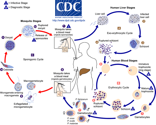
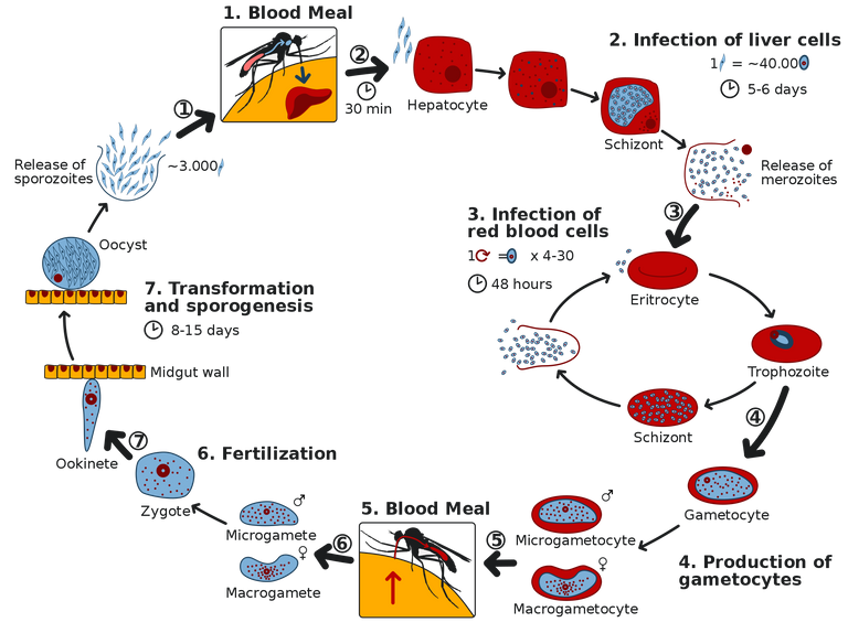
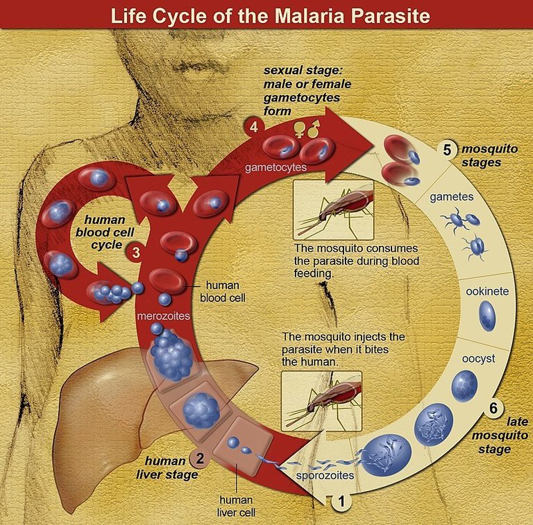
Complete malaria life cycle
Malaria has two life cycle involving an alternating cycles of asexual division (schizogony) occurring in man who is the intermediate host and sexual development (sporogony) occurring in female anopheles mosquito which the definitive host.
During the human cycle which has four stages - pre-erythrocytic (injection of the sporozoites into the blood and its migration to the liver where they transform to schizonts and further into merozoites), erythrocytic (merozoites attack the red blood cells, multiply and release more merozoites that attack other red blood cells. They later transform to trophozoites aka ring form that feeds mostly on the red blood cells, with emphasis on the haemoglobin).
While feeding on haemoglobin, malaria pigments (haemoglozoin) are produced as an end product of haemoglobin breakdown by the parasite. The next stage is
gametogony where the parasite further divides sexually in the red blood cells into - male and female gametocytes. These do not divide further but remain in the blood waiting to be picked up by another mosquito bite in the infected individual. This stage is the last stage known as the dormant or exo-erythrocytic schizogony
In general, mosquito and malaria parasite depend on the availability of blood meal in order to complete their life cycle. In order words, malaria parasites cannot complete their life cycle without humans. Their asexual life cycle is the major part that causes the complications and diseases in humans. Drugs developed are targeted at the various stages of their asexual life cycle in humans.
From the lifecycle we have described above, the schizont, trophozoites (ring form), Malaria pigment, and gametocytes are the major tools for diagnosis. Their presence in the blood film when observed under the microscope determines prognosis. Microscopy remains the gold standard for detection of malaria parasite in a sample and we will critically look at diagnostic procedures shortly.
The life cycle of plasmodium
PS: Most of the above are merged excerpts from my previously written articles about malaria as a disease and the causative agent but with some modifications. For the complete and detailed write-up, you can check them out here - The vampires of the tropics: an exhaustive overview and this - Plasmodium falciparum: the major virulence factor in the pathogenesis of malaria. Both posts practically has all the details you would ever think of. Our main focus is on the practical clinical significance and diagnosis of malaria.
Malaria diagnosis: the diagnostic procedures
Let's clear up something here, so that we don't ever get to mix them up. Mosquitoes are vectors or agents that carry plasmodium. Plasmodium is the pathogen can causes the disease. Now the name of the disease is malaria. This is as clear as it can be.
When carrying out diagnostic procedures to detect malaria parasite using microscopy, schizont, trophozoites (ring form), pigments and gametocytes are the major features looked out for. There presence is what determines if a patient is suffering from malaria or not.
Diagnosis of malaria disease begins with the preparation of the blood film of the patient, and this blood film could either be a thick or thin film. It all depends on choice, but the most favoured for malaria diagnosis, is thick film.
The reason behind thick being preferred is that, thick film contains higher concentration of the parasite. It enables you visualize even the smallest concentration of the plasmodium parasite in the patient sample of blood. With thick film, the chances of seeing a malaria parasite is higher than when compared to using a thin film.
Preparation of thick blood film is pretty simple and straight forward. All you need do is to place a drop of blood at the center of a clean grease-free slide. You then use an applicator stick to spread the blood by making gradual circular pattern of it. Allow it to dry well afterwards before you stain using any Romanowsky stain.
For thin film procedure and staining technique for both, you can read up the 'diagnostic procedure' section of this post - Medical laboratory workshop episode 7: what does differential white blood cell count say about your health condition, you sure will have a better understanding.
Expected results and findings
In diagnosis of malaria, microscopy remains the gold standard, reason being that, most new methods do not accurately identify the plasmodium. The use of microscope correctly reveals the plasmodium parasite in the blood and also reveals the characteristics and forms of these parasite. We will discuss all the forms of this parasite as seen under the microscope.
The commonly seen forms of the plasmodium is the Trophozoites (ring form). Unfortunately, digital microscope is not available at moment, so we will make do with our regular compound binocular microscope as usual. We will only reveal the ones we are able to see and then supplement others with those taken with digital microscope. Let's begin with trophozoites and progress with others.
Trophozoites
The trophozoites of plasmodium are the commonly seen evidences of malaria parasite infection in patients. It has two major very important diagnostic markers - the rind of cytoplasm and chromatin dots. In severe cases, they are always seen numerous in the blood film.
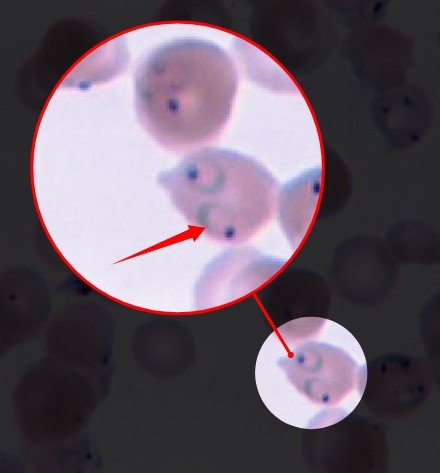 Pointed structure: Cytoplasmic ring of plasmodium falciparum parasite fully attached to the chromatin dot
The cytoplasmic ring is always seen intact and attached to the chromatin dot in cases where the patient has not taken any medication to treat the malaria infection. When the sample preparation procedure is correctly followed, you also tend to see the cytoplasmic ring completely intact and attached to the chromatin dot.
Pointed structure: Chromatin dots of plasmodium falciparum
There are situations that could cause or result to missing cytoplasmic ring. One of the reasons is drugs. When patients take drugs prior sample collection, the chances of seeing the ring still intact will be quite slim because drugs tend to destroy this ring. Secondly, you could loose the cytoplasmic ring during the preparation of the thick or thin film.
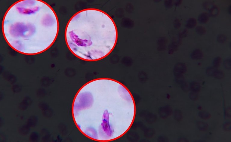
Note that, the chromatin dots in most cases are one, but there are situations where they could be two in trophozoites and still attached to a a common cytoplasmic ring. Depending on the colour of the chromatin dots, experience has shown that you can easily tell if it an early or late infection. Those one that stains light red or pink are as a result of recent and fresh infection while those that stain thicker and deeper in colour (purple-blue) are older infection.
Their location can also vary. Some are seen attached to the circumference of the blood cells. Such type are referred to as the accule form of malaria parasite.
Pointed structure: Accolle form of plasmodium falciparum
You could also see the bacillary form of malaria parasite. They have a rod shaped chromatin dots. In essence, not all chromatin dots are circular in shape. Having this at the back of your mind will help you in easily identifying any form of malaria parasite trophozoites. Having said all the above, let's look at the gametocytes.
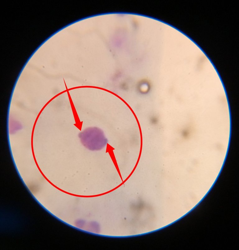
Gametocytes
Gametocytes are another very important diagnostic markers in identification of forms of malaria parasite. They are not often seen as trophozoites.
Highlighted structures: Gametocytes of plasmodium falciparum
They have a characteristic sausag shape or comma with an almost centrally placed pigment that stains darker. Try not to confuse them with monocytes (a type of white blood cell that has similar shape as bean).
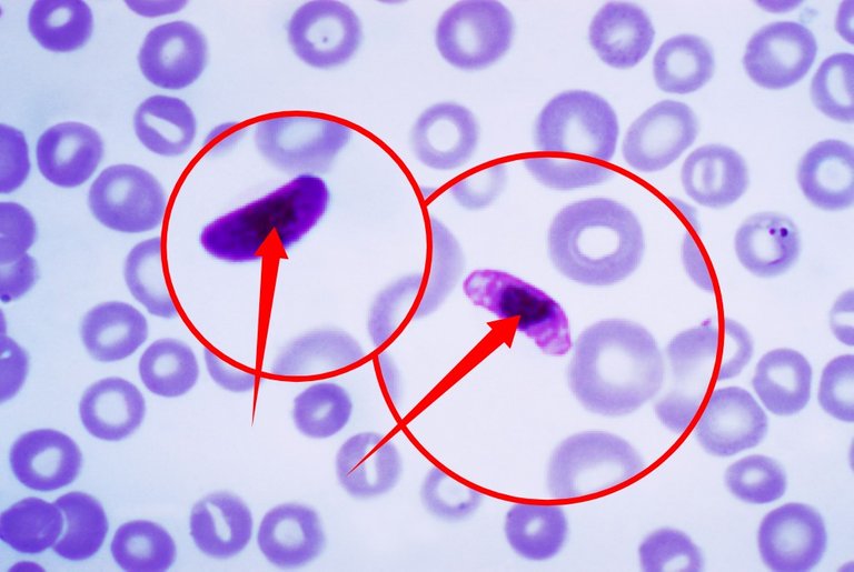
Pointed structure: Centrally placed nucleus of Gametocytes
The major difference between monocytes and gametocytes, lies in the presence of centrally placed dark pigment in gametocytes. Secondly, gametocytes are always smaller in size compared to monocytes which are far larger. Next is the malaria pigments and schizonts.
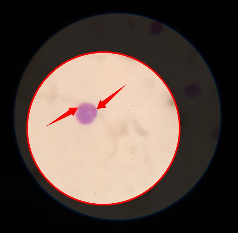
Malaria pigment and schizonts
Just like we earlier explained, malaria pigments are waste products of the degradation of haemoglobins in the red blood cells by the malaria parasite. They are mostly seen in cases of chronic infection with the parasite.
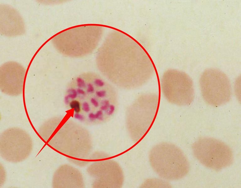
Highlighted Enlarged structure: malaria pigment (brown) within a schizont
The pointed structure in the image directly above is the malaria pigment found within the schizont. Note must be taken to confuse the malaria pigments with chromatin dots. Chromatin dots are smaller in size when compared to malaria pigments which are larger. The pigments stain darker than chromatin dots.
Conclusively, a good understanding and the ability to accurately diagnose malaria disease solely depends on the accurate identification of the various forms of these parasites under the microscope.
Malaria is one of the deadliest disease in the tropics, the vectors of the parasite - mosquitoes must at all times be eliminated by any means necessary. Though efforts have been made to eradicate them, as an individual, you still have a huge role to play in achieving a zero malaria goal. Join the fight by using mosquito nets when need be and also use physical force any means at your disposal.
Until I come your way, stay awesome!
References
•DPDx - Laboratory Identification of Parasites of Public Health Concern
•Comparison of the Plasmodium Species
•Differences Between Plasmodium falciparum and Plasmodium vivax
•How to identify the type of malaria on a blood smear
Thanks for your post.
This is really detailed.
Thanks for the read 💞
My biggest worry every night is mosquito, they are my first problem also when I wake up. If I go to bed and wake up after several bites of mosquitoes, I often wake up feeling sick. I will have a very slight headache, fell weak and a little drowsy. This would happen for a few minutes, sometimes for about an hour, before my body stabilizes. I hope every day that malaria could become history, because it tells on me regularly.
Hopefully it will. Vaccine development is still in its drug trial stage.
In the nearest future, malaria will be a thing of the past.
Congratulations!
✅ Good job. Your post has been appreciated and has received support from CHESS BROTHERS ♔ 💪
♟ We invite you to use our hashtag #chessbrothers and learn more about us.
♟♟ You can also reach us on our Discord server and promote your posts there.
♟♟♟ Consider joining our curation trail so we work as a team and you get rewards automatically.
♞♟ Check out our @chessbrotherspro account to learn about the curation process carried out daily by our team.
Kindly
The CHESS BROTHERS team
Thanks 💞
https://twitter.com/gentleshaid/status/1565397383982333953
The rewards earned on this comment will go directly to the people( @gentleshaid ) sharing the post on Twitter as long as they are registered with @poshtoken. Sign up at https://hiveposh.com.
Thanks for your contribution to the STEMsocial community. Feel free to join us on discord to get to know the rest of us!
Please consider delegating to the @stemsocial account (85% of the curation rewards are returned).
Thanks for including @stemsocial as a beneficiary, which gives you stronger support.
💞