Medical laboratory workshop Episode 2 (A rare occurrence - Live view of Miracidium of Shistosoma haematobium after hatching from egg
In the last episode that birthed this workshop series, we critically explained the life cycle of Shistosoma species and with more emphasis on Shistosoma haematobium which we found in the urine sample of a male patient.
We were also able to establish the various unique features that are associated with the parasite and also we explained the significance of this parasite when they are found in urine or faeces. From the discussion as well, we can conclusively say that, there are only two motile forms of the parasite - Miracidium and Cercariae (the infective form of the parasite).
.png)
Fortunately for us, we were able to see the eggs of this parasite and judging from the flow of the life cycle, the next stage it would form would be the Miracidium. Well, we actually discovered something that has been observed to happen on rare occasion - the presence of a live motile Miracidium within a freshly voided urine sample. Read on, you have one or two things to learn.
This post will be the continuation of the last episode. If you are just joining the series, it would be awesome if you read the previous episode so as to get a good grasps of what we will be discussing. Since the parasite is motile, the best possible way to observe it is through video.
Our pattern of discussion today will slightly be different from last episode. As we discuss, We will as well incorporate our result and findings. Let's briefly learn more about the stage of this parasite called Miracidium.
General overview
Miracidium is the first motile stage of the parasite Schistosoma species and narrowing it down to the specie we saw - Schistosoma haematobium. The Miracidium is released from the egg of the parasite once they come in contact with water. This particular form of the parasite is highly motile.
Note at this point that, the presence of the egg of schistosoma is a good indication of infection with the parasite, but another question that needs to be addressed is, the viability of the eggs. Not all eggs are viable and the easy way to do this, is to carry out manual hatching of the eggs.
How to hatch the eggs of Schistosoma for viability testing
When you hatch the egg of schistosoma and you observe a actively motile Miracidium, then it means the egg is viable. A more direct way to determine the viability of Shistosoma egg, is to do a wet preparation of either the stool sample or urine. The major thing you look out for is, if there is an active moving flame cell in the Miracidium.
First thing first, you need to hatch the egg first before you can do the above viability testing, and to do this, you will need only two things - Normal saline and dechlorinated water (water with no chlorine content). Using water with no chlorine content is necessary because, chlorine has the capacity of killing Miracidia. Hence, if used, it would likely give a false negative result.
The first thing you do is to mix the stool sample or urine sample with normal saline in a glass jar or flask that has a side arm. Allow it to settle for 2 hours or you can spin. For stool sample, you will need to filter the mixture to remove debris. After spinning or allowing to settle for 2hours, you then decant the supernatant fluid.
You then pour the dechlorinated water into the jar and cover the jar with black paper or aluminum foil leaving out only the handle. Keep it near source of illumination over night. After 24hours, use a hand lens to observe the jar, if the eggs are viable, you will see that some have hatched with an active swimming Miracidium inside the water.
Any striking feature of Miracidium?
One remarkable thing about the stage of this parasite is that, it does not feed nor require any source of feeding. Its main focus is on locating any suitable intermediate host so that it can undergo asexual reproduction to produce the infective form, the Cercariae. This asexual reproduction mostly occurs in water snails.
You might want to ask how are they able to locate their host. They do this using light gravity, random motion and chemo sensitivity. Once they find their suitable intermediate host, they penetrate and undergo asexual reproduction as we have explained about and consequently produce the sporocyst form. If they do not find their intermediate host, the chances of survival will be in jeopardy.
Almost all their entire body is covered with cilia of which they use for movement in search of suitable intermediate host. Since they do not feed, they use the store of food within them and if they do not find any suitable host soon enough, they die off. In essence, the availability of intermediate host is very important.
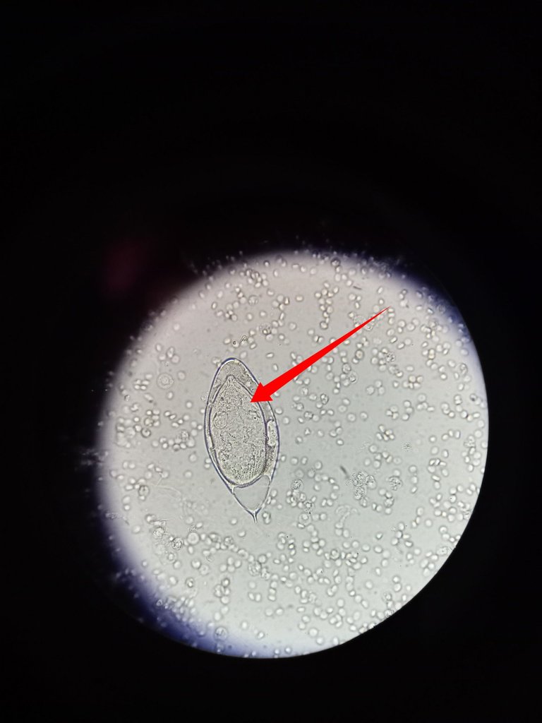
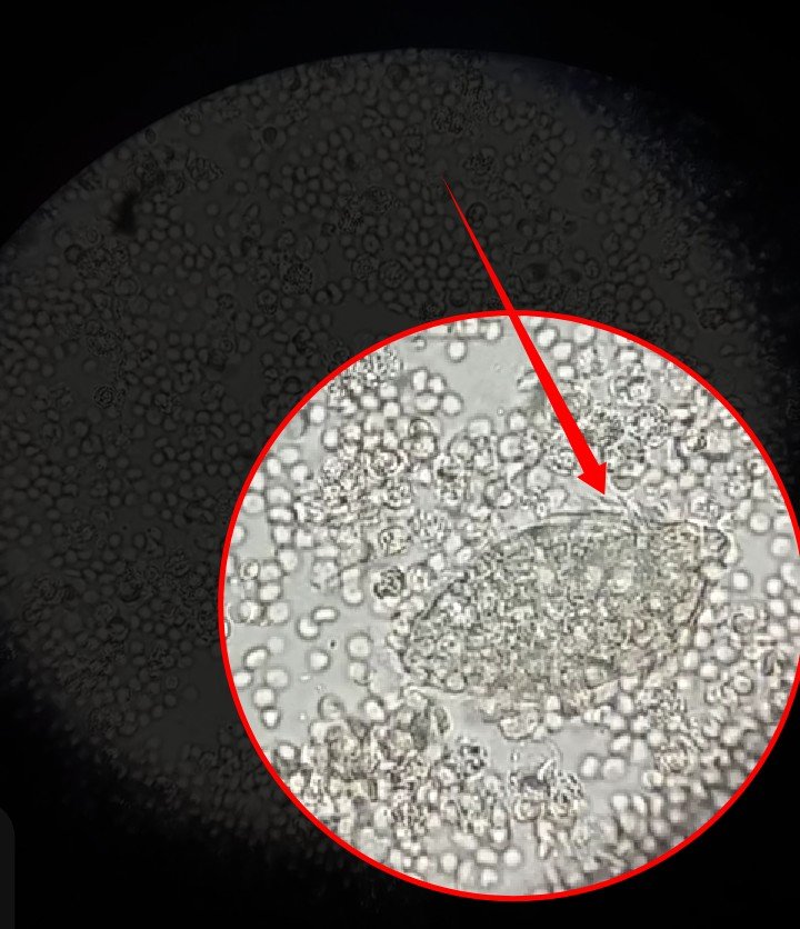
The cilia of the Miracidium are not clearly visible whilst they are within the egg until after hatching. The live image of the captured Miracidium as it moved within blood cells found in the urine of the patient is seen below.
All things being equal, one would expect that this parasite form be found in water or snail only after the egg has been expelled from the body of the infected person through stooling. The case is quite different here. On further critical view of the urine specimen, we were able to discover some hatched motile Miracidium. A nice discovery of which is backed by other report confirming the possibility.
The report of GW Procop and colleagues has shown that their possibility of having miracidium in a freshly voided urine and this has some clinical and diagnostic importance. Though the occurrence has been reported to be rare. Well, we have been able to detect it in our time.
Since this is rare, what then could be the clinical significance of this. Judging from the nature of the boy's urine sample, it had numerous amount of blood, which could be an indication of a chronic infection with this parasite.
Let's briefly discuss look at the significance of the presence of Miracidium in urine sample.
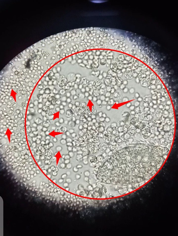
The video below was recorded through the ocular lens of the microscope using an android phone since a digital microscope is currently not available. We had to improvise and I believe it's relatively clear and close enough for visualization of the Miracidium.
For a better view quality, if you are using a mobile device, I would suggest you increase the view by tapping the maximize icon and also change the default YouTube settings to highest quality. For desktop users the latter applies only. Hopefully in time to come, we will get a digitalized microscope.
Watch and observe the parasite as it tries to pass through the numerous blood cells in the field. The movement as can be seen is facilitated by the beating actions of the cilia.
Like we earlier pointed out, Miracidium is expected to be found either in Water or in snail only after the eggs have been expelled from the infected person. When they are present alongside their eggs in the victim, it simply means that the their is an active infection with the parasite and most time would not require egg viability testing.
In this case, it will require urgent treatment to eliminate the parasite before they cause more damage to the bladder. Chronic infection with this parasite has also been reported to be associated with infertility in men. They could also cause bladder cancer.
Treatment of this parasite is not complicated and simply requires administration of Praziquantel. Treatment basically last for a day or two except in cases of complications resulting from the severe damages done by the parasite to the urinary bladder.
Praziquantel helps in the elimination of all forms of the parasite and drug on most trematodes and since Schistosomes is one, it kills it by causing paralysis of the worm's muscles thereby rendering them immobile and unable to feed. They soon die off and are then expelled from the body.
This blog in no way recommends taking the drug without prescription. Ensure you visit the hospital for proper examination.
PS: Aside the first image, others were taken with an Android phone through the ocular lens of the microscope.
References
•Diagnostic value of a miracidium in urinary sediment
•Schistosomiasis
•Schistosoma haematobium
•Hatching of Schistosoma mansoni eggs and observations on motility of miracidia
https://twitter.com/gentleshaid/status/1547539368423866370
The rewards earned on this comment will go directly to the people( @gentleshaid ) sharing the post on Twitter as long as they are registered with @poshtoken. Sign up at https://hiveposh.com.
The world of microbiology is one of the things I am most passionate about, your post is a good material I would say it is a great effort to share with us, thank you for this.
The field is indeed interesting, all thanks to the microscope which enables us to visualize these organisms at high resolution.
Are you using an electron microscope or which one?
Nah, used compound binocular microscope.
Electron microscope is like gold here.
does it have a camera for digital photos?
Not at all. You will need to improvise by using your camera directly (the technique I used to capture the image used here).
Yay! 🤗
Your content has been boosted with Ecency Points, by @cyprianj.
Use Ecency daily to boost your growth on platform!
Support Ecency
Vote for new Proposal
Delegate HP and earn more
Congratulations!
✅ Good job. Your post has been appreciated and has received support from CHESS BROTHERS ♔ 💪
♟ We invite you to use our hashtag #chessbrothers and learn more about us.
♟♟ You can also reach us on our Discord server and promote your posts there.
♟♟♟ Consider joining our curation trail so we work as a team and you get rewards automatically.
♞♟ Check out our @chessbrotherspro account to learn about the curation process carried out daily by our team.
Kindly
The CHESS BROTHERS team
Thanks for the support
Thanks for this great episode. It was quite clear and enjoyable. I however have a (probably very naive) question related to the sentence below, that somehow puzzles me.
How does the parasite produce energy without getting food. I don’t understand this. Its energy must come from somewhere, mustn’t it?
Thanks in advance for clarifying and enlightening my ignorance!
Alright sir,
probably is an oversight.
let me throw more light on it.
Shistosomes mostly feed on proteins and monosaccharides (Glucose) in their host (humans). Their miracidium has a reserve of this nutrient for survival. Once they use up this reserve in them they die-off. This why water and availability of a snails inside water bodies is very crucial for their survival. If they find snail early enough before they use up their energy reserve, they survive, if they don't, that is the end of the road for them.
lovely question sir...
OK I see. So during the time they don't feed, they live on their reserve. I assume thus that they cannot stand very long without a host. Is that correct? Do you have any idea about the time they have?
Cheers!
You are right sir with the duration assumption sir.
I actually didn't know this until now after brief search on the internet...
This is quite a short time to find a host. But where their chances of survival lies is the fact that, the eggs produced are quite numerous.
For example,
While the Shistosoma japonicum lays approximately 2,200 eggs per day
Typical example of the r and K survival strategy. With such large number of egg production, they increase their chances of finding a suitable snail.
Sources - https://www.sciencedirect.com/topics/immunology-and-microbiology/miracidium
https://pubmed.ncbi.nlm.nih.gov/8147487/#:~:text=Schistosoma%20mansoni%20worm%20pairs%20laid,were%20passed%20in%20the%20feces.
That makes sense. Many eggs compensate the smallness of the lifespan. Thanks for having looked for these pieces of information (and the detailed set of references).
Cheers, and have a nice week-end!
Thanks for your contribution to the STEMsocial community. Feel free to join us on discord to get to know the rest of us!
Please consider delegating to the @stemsocial account (85% of the curation rewards are returned).
Thanks for including @stemsocial as a beneficiary, which gives you stronger support.
This is fascinating. I have read about Schistosomiasis before and know that it can be a devastating disease. The video you offer is startling. In watching it we realize that these microscopic life forms have the drive to survive and reproduce, in us. Makes my think of the Guinea worm, another horrible parasite.
Thanks for another great, enlightening article.
Thanks for the compliment and for taking time to go through the work.
Guinea worm akaDracunculus medinensis is Indeed a horrible parasite infection. Especially how it does leave a hole on site of infection.