Cyanotic Congenital Heart Disease Part 2 || Transposition Of The Great Arteries
In my last post, Cyanotic Congenital Heart Disease Part 1 || Tetralogy of Fallot (TOF), I explained the Tetralogy of Fallot (TOF) which is a congenital heart disease with four heart disorders; Ventricular Septal Defect (VSD), Pulmonary Stenosis (PS), Overriding Aorta, and Right Ventricular Hypertrophy (RVH). Today, I will be explaining Transposition Of The Great Arteries, which is another Cyanotic Congenital Heart Disease.
Epidemiology of Transposition Of The Great Arteries
Transposition of the great arteries is the most common cyanotic congenital heart disease with a prevalence of about 2.3-4.7 per 10,000 childbirth., and 5-7% of all cardiac malfunctions.. Patients with TGA usually have associated congenital heart disease. 50% of patients with TGA have ventricular septum defect (VSA).
Embryology of Transposition Of The Great Arteries
In explaining this congenital heart disease, understanding the heart and its circulation is very important. You can read my post Another Child Born With a Heart Defect || Acyanotic Congenital Heart Disease to learn about the structure of the heart but for the purpose of this post, I will be doing a small explanation. The heart is divided into four chambers (2 Atrial, and 2 Ventricles), which are divided into the left Atrium and Left Ventricle, and the Right Atrium and Right Ventricle. The ventricles are responsible for Pumping Bloods (the right ventricle pumps blood to the Lungs, and the left ventricle pumps blood to the body). The Right Ventricle is connected to the pulmonary artery, which takes deoxygenated blood to the lungs as to return oxygenated blood to the left atrium, which then goes to the left ventricle and is pumped to the body..
While the heart functions like that in a baby or a matured human, it doesn't function that way in a fetus. In the fetus, the lungs isn't matured, and there are a lot of pressure on the lungs, so the heart doesn't pump blood to the lungs. In fetal circulation, the fetus gets oxygenated blood from the Umbilical vein (Oxygen rich blood comes from the placenta to the fetus and the mother does the work of the lungs). The oxygenated blood from the Umbilical vein gets into the right Atrium before going to the right Ventricle but in the right Atrium, there is an opening which connects the right Atrium to the left Atrium, known as the Patent Foramen Ovale which allows for the shunting of blood from the right Atrium to the left Atrium. The blood shunt from the right atrium to the left atrium goes to the left ventricle. In the fetal circulation, there is the Patent Ductus Arteriosis, which is a connection between the pulmonary artery connected to the right Ventricle, and the Aorta connected to the Left ventricle in the fetus. Blood going through the Pulmonary Artery bypasses the lungs to the Aorta, and then is circulated to the fetal body. Remember that the two openings allow for the shunting of blood from the right side of the heart to the left side of the heart..
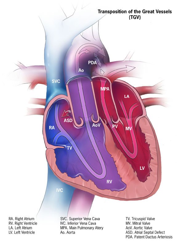
Transposition Of The Great Arteries is a congenital defect that is as a result of the aorta and pulmonary artery are transposed, in that the Aorta is situated in the right ventricle while the Pulmonary artery is attached to the left ventricle, instead of the reverse. The great arteries which are supposed to be crossing one another are parallel to one another as a result of the transposition. The development of Transposition Of The Great Arteries in children hasn't been elucidated, but it is believed that a well established theory is that the condition arises from the abnormal development of the Bilateral Sub-arterial Conus. During the first month of gestation, the sub-aortic conus and sub-pulmonary conus, which serves as the great arteries for the fetus, are located at the right ventricle. In a normal scenario, when the fetus reaches about 34 days of gestation, the Sub-aortic conus is reabsorbed to allow the Aortic valve to be positioned on the left ventricle while the Sub-Pulmonary conus remains attached to the right ventricle thereby allowing the pulmonary valve remain attached to the right ventricle. In the case of Transposition Of The Great Arteries, the case is different. Instead of the Sub-aortic conus to be absorbed in and allow the aortic valve to be positioned in the left ventricle, the Sub-pulmonary conus is absorbed. With this, the Pulmonary valve is then attached to the left ventricle, while the Aortic valve remains attached to the right ventricle.. In a normal condition, the Aortic valve is located in the PosteriorInferior of the left ventricle, but with Transposition Of The Great Arteries, the Aorta is found in the anteriorinferior of the right ventricle.
Pathophysiology of Transposition Of The Great Arteries
With Transposition Of The Great Arteries, the Aorta and the Pulmonary artery are placed in the wrong position (having the Aorta attached to the right ventricle and the pulmonary artery to the left ventricle). With this, when the deoxygenated blood comes from the superior-inferior vena cava, it goes into the right Atrium and then passes the blood to the right Ventricle. The right ventricle is meant to sent the deoxygenated blood to the lungs to get oxygenated but because the Aorta is connected to the right ventricle, the deoxygenated blood goes to straight to the body which leading to cyanosis. For the oxygenated blood, the lungs pumps the oxygenated blood via the pulmonary vein to the Left Atrium. The left Atrium then moves the blood to the left ventricle, which then pumps the oxygenated blood to the lungs back though the pulmonary artery that is attached wrongly to the left ventricle. The pattern of circulations makes it fall under the critical congenital heart diseases category which could be life threatening and requires an immediate and mandatory fixing..
Diagnosis, and Management of Transposition Of The Great Arteries
Before birth, Transposition Of The Great Arteries can be diagnosed using fetal echocardiography. In Postnatal diagnosis, Electrocardiography (ECG), Chest X-ray, Cardiac catheterization and Echocardiography (Echo), will be required..
Managing TGA is considered a critical disease which should be fixed immediately. With that, performing surgery within the first week of life (within 3 - 5 days) is important. The surgery deals with correcting the Arteries. IV infusion of prostaglandin E1 would temporarily maintain the ductus arteriosus. This isn't a solution, surgery is often the required in this case.
Images
Image 1 || Wikemedia commons
Thanks for your contribution to the STEMsocial community. Feel free to join us on discord to get to know the rest of us!
Please consider delegating to the @stemsocial account (85% of the curation rewards are returned).
You may also include @stemsocial as a beneficiary of the rewards of this post to get a stronger support.
Great content. Congenital heart diseases are serious medical conditions and leading causes of infant mortalities especially in resource poor settings where open heart surgeries cannot be easily done.
True.