Neurology Explained - The Oculomotor nerve (The Cranial Nerve 3)
Most times I go out ways to start my post. Some days, I want to say Hi everyone, but that looks old-fashioned, other days, I just want to go straight to the point, but it makes my post look like an ice cream without a topping. That said, I guess I already began the post, I do not have a reason to worry about how to start it anymore. Over the last few days, I have been going through what you can call a series, a course, or some people may even call it a sequence on the cranial nerve, pardon my use of English if it doesn't meet your standard.
English can be very brain stressing for me, even when it is the L1 language (Lingua franca), of my country. Anyway, you would have you endure with me on this some worth rusty ride. Okay! Now back to business.
I explained the first and second cranial nerve, which are the olfactory nerves and the optic nerve respectively. If you missed them, you should do well to check them out on my blog. I could be nice enough to also put the links at the end of the post, I am sure that was a relief for you. [Just do well to read them because I could also forget to put at them at the end of the post. Anyone could suffer from 5 minutes partial amnesia :)].
Today, I will be looking into the third cranial nerve (Cranial Nerve III) which is the Oculomotor nerve. Remember in my last post, I made it clear that the Optic Nerve isn't the only nerve responsible for innervating sight, as there are more than one nerve responsible for it. Today, I will be explaining the Oculomotor nerve.

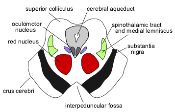
The Brain Stem and the Oculomotor nerve Nucleus
You might have possibly heard that the nucleus of the oculomotor nerve is found within the midbrain of the Brain stem, and that is true, but let me quickly do a little neuroanatomy of the midbrain. The midbrain has in its center, the Cerebral Aqueduct which contains cerebrospinal fluid, superiorly connected to the third ventricle of the brain, and inferiorly connected to the fourth ventricle of the brain. Just beside the cerebral aqueduct, is the nucleus of the oculomotor nerve (cranial nerve III). Away from the oculomotor nerve nucleus, by the sides, are the Edinger-Westphal Nucleus, which are parasympathetic nucleus. Anterior to the oculomotor nerve (cranial nerve III) is the Crux Cerebri, at the sides of the Edinger-Westphal Nucleus are the Medial Lemniscus which has the spinothalamic tract right above it (posteriorly). Posterior to the spinothalamic tract, are the superior colliculus (remember in my last post I mentioned that the Superior Colliculus is responsible for the control of reflexive eye movement). Interior to the medial lemniscus is the Red Nucleus. After my very brief anatomical explanation, it would be perfect to say that posterior to the nucleus of the Third Cranial Nerve (Cranial Nerve III), which is the Oculomotor Nerve, is the superior colliculus, beside are the medial lemniscus, and anterior to it are the red nucleus. The cranial Nerve fibers (somatic motor fibers) of the oculomotor nerve, along with the parasympathetic nerve fibers, move through the red nucleus out of the interpeduncular fossa.
In the brain stem, the Cranial Nerve III along with the parasympathetic nerve fiber will run between the Posterior Cerebral Artery and the Superior Cerebellar Artery, with the movement of the Cranial Nerve III runnung under the Posterior Cerebral Artery and above the Superior Cerebellar Artery, and the runs through the lateral wall of the Cavernous sinus and go into the superior orbital fissure. At the end of the superior orbital fissure are two arterial branches, the superior and inferior branch. In the inferior branch, the oculomotor nerve moves into a ganglia (cell bodies), known as Ciliary ganglion, which is a postganglionic motor neuron. The ciliary ganglion through ciliary nerves supplies to the sphincter pupil and the ciliary muscle which causes the expanding and retracting of the lens for near and far vision., . At the superior branch, the oculomotor nerve connects to the superior tarsal plate close to the levator palpebrae superiors, and the superior rectus.
For ease of knowing which of the nerves control which of the eye muscles, you should remember the (LR6, SO4, and ATR3) which mean that the Lateral rectus is control by the 6th cranial nerve, the Superior oblique is controlled by the 6th cranial nerves, while all other muscles are controlled by the 3rd cranial nerves.
The Third Cranial Nerve has the inferior branch, the superior branch and the parasympathetic nerve fibers (this is the nerve that has been with it since its origin in the brain stem). To start with, the parasympathetic nerve fibers controls the iris, pupil size (which controls the amount of light entering the eye by adjusting the Sphincter pupillae to adjust the pupil), ciliary muscle, and the lens., . The Superior Branch of the oculomotor nerve controls the superior rectus (elevating the eyeball), and the levator palpebrae superiors (elevating the upper eyelid).. The inferior branch of the oculomotor nerve controls the inferior rectus, inferior oblique and the medial rectus..
Conclusion
The third cranial nerve originates from the Midbrain, at the level of the superior colliculus, which is moved with the General somatic efferent fibers, and the Parasympathetic nerve fiber (GVE). It is responsible for supplying the majority of the eye muscles, which I listed above, and the Third cranial nerve is responsible for the eye and eyelid movement.
Image 1 || Flickr || Eye Cranial Nerve
Image 2 || Wikimedia Commons || Midbrain superior colliculus
Yay! 🤗
Your content has been boosted with Ecency Points, by @eni-ola.
Use Ecency daily to boost your growth on platform!
Support Ecency
Vote for new Proposal
Delegate HP and earn more
This is well articulated.
You did a nice job here
Thanks a lot for the time. I am glad you did spent time on my post.
Sincerely, Your posts are always detailed. It takes extra research to get these details and when I read your post and click on the links, they lead me exactly to what you are saying. I need to learn how you write these constructive posts and how you take your time to research a lot of these things. I must confess that this is a post well written. Thanks for sharing.
Haha... I just love doing a lot of research.Like to dig into everything I can dig into, including food.😄😄😄😄
Thanks for your contribution to the STEMsocial community. Feel free to join us on discord to get to know the rest of us!
Please consider delegating to the @stemsocial account (85% of the curation rewards are returned).
Thanks for including @stemsocial as a beneficiary, which gives you stronger support.
Glad to see you are persevering on that topic! I am looking forward to the next episode ;)
Thanks for always checking my post. I am glad you do regularly. I will keep on the series.
I am trying not to miss any of them, but this is not easy. I now follow quite a bit of authors, so that missing one or two posts becomes likely. However, I always come back ;)
A great post, really had me thinking and my eyes blinking 😂
Haha!! The eye is complex, and we really do not take into consideration its complexity and fragility when considering its care. I hope you blink less :)
No problem, it was an interesting post