Neurology Explained - The Vestibulocochlear nerve (Cranial Nerve VIII) || The Vestibular Pathway
It is another day to continue on the Cranial Nerve VIII (Vestibulocochlear nerve). In my last post, I discussed the Auditory pathway, which looks into the process of hearing, but in this post, I will be looking into the vestibular pathway. For me to get to this point, for proper understanding, I decided to create a lot of post to help you. If you reading this post and want to understand it properly, then you should read the posts I wrote on the anatomy of the ear and the inner ear, the cochlea, the post on the semicircular canals, the vestibules, and as well my previous post, the auditory pathway of the vestibulocochlear nerve. Today I will be discussing about the vestibulococlear nerve, and in this post, I will be looking into the vestibular pathway.
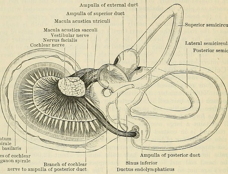
From my previous post, I made it known that there are two pathways in the vestibulocochlear nerve, and that's the auditory/cochlear branch with deals with hearing and its mechanism from the spiral of corti, while the other pathway deals with balancing (remember the vestibular post I made and how the deal with equilibrium?). Yes! so for clearity, the nerves from the cochlea, and the nerve from the vestibules (ampulla, maccula, utricle, and semicircular duct) is what makes up the vestibulocochlear nerve.
Nerves from the vestibules or the vestibular nerve moves through the internal acoustic meatus in the skull, remember I mentioned this in my previous post that the internal acoustic meatus accepts both the cochlear branch, and the vestibular branch, along with other nerves;
The Cochlear nerve fibres goes through the internal acoustic meatus to reach the Pontine-medulla (pontomedulla) junction. Other nerves that go through the internal acoustic meatus are the vestibular nerve (to form the vestibulocochlear nerve), facial nerve (CN VII), and the labyrinthine artery. - Neurology Explained - The Vestibulocochlear nerve (Cranial Nerve VIII) || The Cochlear (Auditory) Pathway
The vestibular branch then moves into the medulla, and the pons-medulla junction. In the medulla, it reaches the vestibular nuclei (bilateral). In the nucleus of the vestibular nerve, there are four sub nuclei divisions, and these are the Lateral vestibular nucleus (LVN), Medial vestibular nucleus (MVN) also known as nucleus of Schwalbe, Superior vestibular nucleus (SVN), Descending (inferior) vestibular nucleus (DVN).. The lateral and the medial vestibular nucleus are responsible for the actions in the descending motor pathways. Nerves from the lateral nucleus and medial nucleus descends via axons known as the vestibulospinal tract through the ventral part of the lateral funiculus, after which it moves to the ipsilateral ventral horn., . The vestibulocochlear nerve then branches to the ipsilaterally to activate the alpha neurons in the lower motor neuron, to move to extensor muscles of the back, knee and extremities, as well as the head and neck muscles (medial vestibular nucleus) for the purpose of resisting gravity and for posture maintenance. ,.
The inferior and Superior vestibular nucleus has its axons which moves into the cerebellum through the inferior cerebellar peduncles. In the cerebellum, it goes into the flocculonodular lobe also known as the vestibular cerebellum. The branch in the cerebellum goes to another nucleus known as the Fastigial nuclei which is responsible for balancing posture, modulating vestiblar spinal nucleus, and maintaining reflexive eye movement.,.
The medial vestibular nucleus sends an axon across the contralateral abducens nerve nucleus, where the axons are sent to different nerves nucleus including the contralateral paramedian pontine reticular formation (PPRF) and contralateral motor nuclei, of the third cranial nerve and the forth cranial nerve nucleus, to form the medial longitudinal fasciculus. These supplies the muscles of the lateral rectus (6th Cranial nerve), Medial Rectus (3rd cranial nerve), and the 4th cranial nerve (trochlear nerve) supplies the superior oblique. The medial vestibular nucleus sends these axons to perform the function of eye fixation during gaze and movement in horizontal or vertical movement. the Vestibular Nucleus also feed the Paramedium pontine reticular formation for the purpose of maintaining gaze when moving or rotating the head.,.
The superior vestibular nucleus also moves upwards, to the ventral posterior medial nucleus also known as the ventrobasal complex, in the thalamus, supplying the insular cortex, temporal perietal cortex, and lateral post central gyrus, for the purpose of creating awareness about the vestibular sensations that occurs in the body. ,,.
Conclusion
The vestibular pathway of the vestibulocochlear nerve is completely different in supplies and pathway from the cochlear pathway. While the both axons enter into the inter acoustic meatus together, the vestibular axon moves to the vestibular nuclei where each of the nucleus send axons to different muscles, and regions. including the cerebellum, the cortex of the midbrain, the upper medulla, and downwards through the vestibulospinal tract to the extensor muscles of the back, knee and extremities. While the cochlear nerve of the vestibulocochlear nerve is for auditory purposes, the vestibular nerve is responsible for maintaining balance, and equilibrium.
Image Reference
Image 1 || Flickr Image || Cunningham's Text-book of anatomy
Thanks for your contribution to the STEMsocial community. Feel free to join us on discord to get to know the rest of us!
Please consider delegating to the @stemsocial account (85% of the curation rewards are returned).
Thanks for including @stemsocial as a beneficiary, which gives you stronger support.