The Anatomy of the Facet Joint, and Facet Joint Dysfunction
Brief Introduction
There are numerous joints in the body. I am sure in elementary school, you were taught the movable joints. When I was in elementary school, I remember my teacher will always state the movable joint and would mention the bow and socket joint, the Pivot joint, the hinge joint, and the gliding and sliding joints. These joint listings were all I believed were movable joints. Anyways, in this post, I will be discussing the Facet Joint in the spine today you should get ready as we explain in detail everything you should know about the Facet joint.
Facet Joint Simple Anatomy
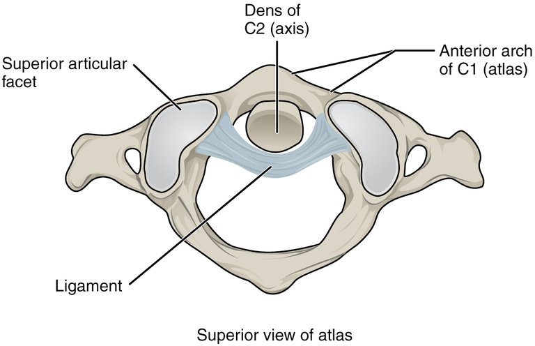
The vertebral column is made up of two adjacent vertebral bodies, an intervertebral disc, and two facet joints. The Facet joint is a type of synovial joint also known as the zygapophyseal joint which is an articulation between the inferior articular facet of the vertebrae and the superior facet of the vertebrae. It is located between the pedicle and the lamina of the vertebrae. The Superior articular facet is oriented superior and posteriorly (it is on the vertebrae below facing upward), while the inferior articular facet is oriented inferiorly and anteriorly (it is on the vertebrae above facing downwards). Both articular facets are being held by the facet joint capsule. The joint is oriented at an angle of 45 degrees horizontally, and both facets are lined by the hyaline cartilage.
Quick explanation
Synovial joints are a type of movable joint found throughout the body, characterized by the presence of a synovial cavity, a fluid-filled space between the articulating bones.
Hyaline cartilage is a smooth and glossy connective tissue made up of collagen and proteoglycan. it is found in the bone of joints, the ear, the nose, and the trachea, acting as a shock absorber in these areas.
commons.wikimedia.org
The facet joint is made up of a fat pad which serves as a cushion in the joint. These fat pads aren't bones, they are adipose tissues with blood running through them as well as have a nerve supply, giving them the ability to bleed with trauma, as well as cause pains since they can send potential pain sense through their nerves. The facet joint is innervated by the medial branch of the Dorsal Ramus of the spinal nerve, and irritation to the facet joint would lead to the release of inflammatory mediators, irritating the nerve root.
Quick explanation
- The dorsal ramus is a spinal nerve branch that originates from the spinal cord and moves towards the posterior (dorsal) side of the body, supplying motor and sensory innervation to the muscles, skin, shoulder, upper arm, and back muscles, as well as supply sensation to the skin of the posterior trunk and limbs.
Facet Joint Dysfunction
The Facet joint is always filled with synovial fluid and stored by the synovial membrane. Its function is to lubricate the joint, while acting as a shock absorber and helping to reduce friction between both articulating bones. The facet joint allows for the flexion and extension of the spine, and prevention of the vertebrae from slipping over themselves. When the facet joint, and its associate experience trauma causing it to become inflamed, or damaged, resulting in a source of pain, then it is regarded as facet joint disease or syndrome or arthritis. There are different types of facet joint disease associated with its pathology, but Facet osteoarthritis is the most common type of facet pathology.
There is 65 to 80% of average American adult suffers from Loa back pain, and it is at a consistent prevalence level as a result of degeneration of the joints, and the bones over time. A high-risk factor for having Facet joint Osteoarthritis is a history of heavy work before the age of 20 years. Cervical facet disease has a prevalence of between 29% and 60% of cases, and neck pain also is common, and has become a leading contributor to disability over the years. Study shows that 30% to 50% of neck pains become chronic after 12 months of their prevalence.
Etiologies of facet joints
Facet joint effusion
This condition occurs as a result of excessive fluid buildup in the facet joint as a result of an injury, degeneration, or inflammation of the facet joint. Facet joint effusion can occur as a result of degenerate spondylolisthesis, spinal instability, other degenerative disc diseases, or a herniated disc.Facet joints synovial cysts
Facial joint synovial cysts are a condition associated with fluid-filled sacs which develop in the facet joints as a result of degeneration or inflammation of the facial joint causing the fluid to create pressure inside the spinal canal compressing the nerves. Its pathogenesis remains unclear and is regarded as a degenerative disorder but it can also be a result of spinal instability and trauma.Osteoarthritis of the facet joints
Osteoarthritis of the facet joints is degenerative arthritis affecting the small synovial joints of the spine. it usually begins as a degenerative change in the articular cartilage and extends to the synovium, joint capsule, subchondral bone, ligaments, and musculature. It can be caused by the wear and tear of the cartilage leading to inflammation causing pain and stiffness. It is common in elderly people with injury or other forms of Arthritis.Rheumatoid arthritis
Rheumatoid arthritis (RA) is a chronic autoimmune disorder that causes inflammation, and damage to the joints, causing pain. In this condition, the synovium is attacked leading to inflammation and causing fibrovascular proliferation (pannus formation). It can also attack the knee, hips, and shoulders, leading to bone erosion as well as ligamentous laxity.Spondylolisthesis
Spondylolisthesis is a degenerative condition that leads to the displacement of one vertebra over the other in the sagittal plane. This can occur as a result of various reasons including a stress fracture, degeneration of the joints, and congenital defects, causing pain, weakness, numbness, and progressive loss of cartilage and articular remodeling.
Symptoms and Evaluation of Facial Joint Dysfunction
Symptoms include sharp, localized, and unilateral pain in the area of inflammation, or degenerative facet joint. Patients could experience spasms in that area, Neck and back pain, and disc herniations. Clinical Evaluation would include checking if patients have neck or back pain, and the use of imaging such as MRI, CT, and X-ray to show degeneration, narrowing, hypertrophy and calcification of the joint, and tenderness upon palpation.
Treatment and Management
Conservative management is the first line of therapy to treat Facet Joint Dysfunction. Anti-inflammatory medications are given to treat the mediated pain, weight loss is advised, use of muscle relaxers, physical therapy, and massage are therapies that can be used. When changes are not visible with conservative measures, interventional procedures would be considered, use of radiofrequency ablation
- National Library of Medicine - Anatomy, and pathology of facet joint
- Dovepress.com - Facet Joint Syndrome: Pathophysiology, Diagnosis, and Treatment
- National Library of Medicine - Facet Joint Disease
- National Library of Medicine - Facet joint syndrome: from diagnosis to interventional management
- Radio Graphics - Differential Diagnosis of Facet Joint Disorders
- Physio-pedia.com - Facet Joint Syndrome
- National Library of Medicine - The human lumbar dorsal rami
I believe that joint pain are always very severe even with the slightest of injury, the connection of neurons in the joints to other body part gives that strong sensational feeling (I stand corrected), thanks for producing such educational content.
Yay! 🤗
Your content has been boosted with Ecency Points, by @oluwatobiloba.
Use Ecency daily to boost your growth on platform!
Support Ecency
Vote for new Proposal
Delegate HP and earn more
Thanks for your contribution to the STEMsocial community. Feel free to join us on discord to get to know the rest of us!
Please consider delegating to the @stemsocial account (85% of the curation rewards are returned).
Thanks for including @stemsocial as a beneficiary, which gives you stronger support.
Very interesting
!1UP
You have received a 1UP from @gwajnberg!
@stem-curator, @neoxag-curator
And they will bring !PIZZA 🍕.
Learn more about our delegation service to earn daily rewards. Join the Cartel on Discord.