Pathophysiology of Avascular Necrosis (AVN)
Hello Hivers!
In today's article, we'll talk about Avascular Necrosis. Avascular necrosis is focal bone infarction, which may be secondary to various conditions or idiopathic. Severe osteoarthritis can result. Symptoms include bone or joint pain. Early diagnosis is best made by MRI. Treatment includes rest, analgesics, range-of-motion exercises, and, in some cases, surgery.
Let's go.
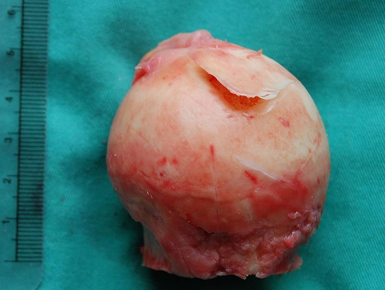
The head of the femur. By Steven Fruitsmaak - Own work, CC BY-SA 3.0.
Avascular necrosis is basically the death of bone tissue due to a lack of blood supply.
In the US, avascular necrosis (AVN) affects about 20,000 new patients annually and causes 5% of cases of osteoarthritis of the hip. The femoral head (hip) is most commonly involved; the distal femur and proximal tibia (knee) and the humeral head (shoulder) are frequently involved; the bodies of the talus, carpal, scaphoid, and navicular bone are less commonly involved. In 33 to 72% of patients with non-traumatic AVN, the disease is bilateral. Idiopathic AVN of the hip affects men 4 to 5 times more often than women, with a peak incidence between ages 30 and 60. AVN of the knee is most common in elderly women.
Causes
AVN involves ischemic death of osteocytes and other bone marrow components, producing subchondral bone infarction. Of the many conditions associated with AVN, some are more clearly causative.
The causes may be definite or probable. Definite causes include alcohol abuse, coagulation disorders, corticosteroids (high dose), decompression sickness, fatty liver, fracture of the femoral neck, Gaucher's disease, hip dislocation, radiation therapy, sickle cell disease and tumours.
Probable causes include atherosclerosis, Cushing's syndrome, diabetes mellitus, gout, Legg-Calvé-Perthes disease, lipid disturbances, pancreatitis, SLE and smoking.
Posttraumatic AVN develops when blood supply is impaired. Susceptible bone is usually intra-articular and has a limited attachment of soft tissue and accompanying vasculature. The hips, shoulders, body of the talus and carpal scaphoid are commonly affected.
AVN after a hip dislocation or fracture results from tears of the ligamentum teres and joint capsule that compromise blood vessels. Posterior dislocations are especially likely to result in AVN. AVN develops in 52% of hips that remain dislocated for greater than 12 hours but in only 22% of those reduced within 12 hours. AVN after hip fractures is common with transcervical or subcapital fractures but is rare with intertrochanteric or other extracapsular fractures. The incidence does not seem related to the surgeon's skill or type of fixation device.
The most common risk factors for nontraumatic AVN are alcohol consumption, use of greater than 25 mg/day of prednisone for several months or more, and sickle cell disease. Less common risk factors include decompression sickness, Gaucher's disease, tumours (lymphomas, leukaemias, metastatic bone tumours), vascular disease (including vasculitis), and radiation therapy.
About 25% of cases occur in patients with no detectable risk factors and are considered idiopathic. However, some patients have clotting abnormalities due to protein C or S deficiencies, hyperhomocysteinemia, or anticardiolipin antibodies.
Signs and Symptoms
Although pain may develop insidiously, many patients remember the hour when they first felt incapacitating pain. As the bone progressively collapses, almost all patients develop pain aggravated by mechanical stresses (e.g., in the hip; standing, moving, walking) and eased by rest. Eventually, 67% of patients have pain at rest and 40% have pain at night, which may be accompanied by prolonged morning pain and stiffness.
AVN of the hip produces groin pain that intermittently radiates down the anteromedial thigh. Some patients note increased pain and a distinct clicking with motion, particularly when rising from sitting. They have limited motion (particularly flexion, abduction, and internal rotation) and adopt an antalgic gait. A click may be elicited by externally rotating the flexed abducted hip (Figure 4 sign).
AVN of the knee usually produces sudden pain without preceding trauma. It is usually localized to the medial side of the knee. There is tenderness over the medial femoral condyle and, in 1⁄3 of patients, mild to moderate effusion.
AVN of the shoulder may produce only transient and minimal symptoms until advanced disease develops. The major complaints are pain and limited motion, particularly active motion. Passive motion is preserved. It is unusual for AVN to involve just the shoulder.
Diagnosis
After fractures, increased pain and limp or deterioration of joint function suggest AVN. Early diagnosis requires a high index of suspicion in patients with conditions associated with AVN who develop pain in their hips, knees, or shoulders.
Diagnosis is made by an imaging study. Plain x-ray is usually performed first but is insensitive and can be normal for months or even years after the onset of symptoms. Early x-ray changes include subtle osteosclerosis and osteoporosis. The femoral head may develop subchondral lucency (the crescentic signal). Next, bone collapses and degenerative joint changes occur.
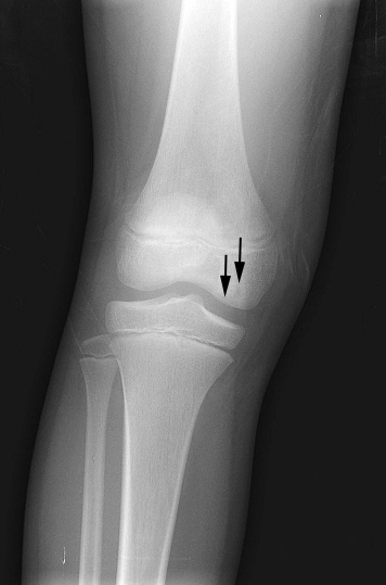
Front X-ray of the right knee of an adolescent (epiphyseal plates are open): arrows point to avascular necrosis and developing osteochondritis dissecans in the outer medial condyle of the femur. By Pil Kang, Affiliation: Uniformed Services University - Walter Reed Army Medical Center. Obtained from MedPix Database, Public Domain.
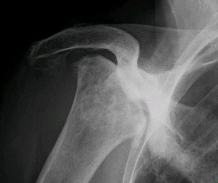
Radiography of total avascular necrosis of right humeral head. Woman of 81 years with diabetes of long evolution. By Jmarchn - Own work, CC BY-SA 3.0.
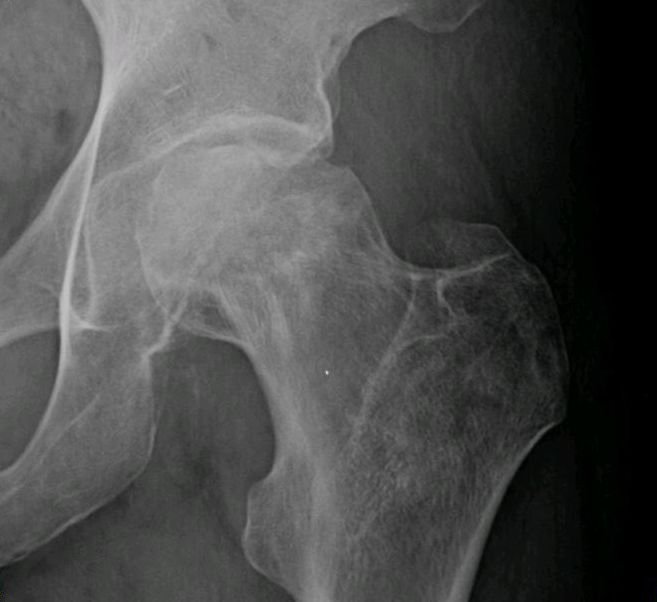
Radiography of avascular necrosis of left femoral head. Man of 45 years with AIDS. By Jmarchn - Own work, CC BY-SA 3.0.
MRI is the most sensitive and specific test. Bone scans also are sensitive and accurate and can screen multiple joints simultaneously. A CT scan may be needed to gauge the degree of bone collapse, which may predict the response to treatments such as surgical core decompression.
Prevention and Treatment
To minimize AVN from corticosteroid use, patients should be given as low a dose as needed for effect and for as short a duration as acceptable. To prevent AVN from decompression sickness, patients should adhere to decompression tables during deep-sea diving.
Conservative measures often include analgesics, range-of-motion exercises, and restriction of weight-bearing (if knees or hips are affected), in an attempt to prevent or minimize bone collapse. Failure of conservative treatment is usually apparent within 2 years. Patients should be followed for at least this long.
Early conservative surgical intervention with core decompression or cortical bone grafts may best prevent serious joint dysfunction in the hips and knees, but surgery is less often necessary in the shoulder. Surgery is most successful if done before the bone collapses.
Core decompression involves drilling a core of bone out, which reduces intraosseous pressure and promotes revascularization.
Electrical stimulation (subthreshold) has been used in conjunction with core decompression to stimulate new bone formation and prevent collapse. If performed before the radiographically detectable collapse, up to 75% of patients avoid joint replacement. Cortical bone grafts may provide mechanical support to prevent collapse as the bone revascularizes. Because this is a more extensive procedure than core decompression, crutches must be used for 4 to 6 months as compared to 2 to 3 months for core decompression.
Osteotomy might also be used to alter mechanics of the joint and redistribute the maximal load-bearing forces to prevent collapse and deformation. Crutch walking is required for 6 to 12 months.
If joint abnormalities develop, severe degenerative changes are inevitable. If the pain becomes severe and is not relieved by nonopioids, total joint replacement is indicated. Because prosthetic hips and knees last 15 or 20 years, total joint replacement is often the procedure of choice for patients greater than 65, whose prosthesis may last for the rest of their life. Younger patients, especially those who persist in vigorous physical activities, will likely need a second procedure.
Do you have any questions, concerns or anything to add? Tell me what you think below.


Resources
- Khan AN, Al-Salman MJ, Chandramohan M, MacDonald S, Hutchinson CE. "Bone Infarct". eMedicine Specialties. Archived
- Chapman C, Mattern C, Levine WN (November 2004). "Arthroscopically assisted core decompression of the proximal humerus for avascular necrosis". Arthroscopy.
- Fujisawa Y, Masuhara K, Shiomi S (1979). "The effect of high tibial osteotomy on osteoarthritis of the knee.
- Mary Anne Dunkin. Avascular Necrosis (AVN or Osteonecrosis) [Internet]. WebMD. WebMD; 2010. Available from: https://www.webmd.com/arthritis/avascular-necrosis-osteonecrosis-symptoms-treatments
- Barney J, Piuzzi NS, Hossein Akhondi. Femoral Head Avascular Necrosis [Internet]. Nih.gov. StatPearls Publishing; 2019. Available from: https://www.ncbi.nlm.nih.gov/books/NBK546658/


Written by @gamsam - a Medical Student
All images used are copyright free
Vancouver Style was used for References.
Thanks for your contribution to the STEMsocial community. Feel free to join us on discord to get to know the rest of us!
Please consider supporting our funding proposal, approving our witness (@stem.witness) or delegating to the @stemsocial account (for some ROI).
Thanks for using the STEMsocial app and including @stemsocial as a beneficiary, which give you stronger support.
test !BEER
Sorry, you don't have enough staked BEER in your account. You need 24 BEER in your virtual fridge to give some of your BEER to others. To view or trade BEER go to hive-engine.com
test !BEER
You cannot sent token to yourself.